


Diabetes

What is Diabetic Retinopathy?
Diabetes is a condition which affects the body's ability to use and store sugar. Incidence of the disease is reaching epidemic proportions in our country—increasing six-fold in the last 30 years. Medical achievements have enabled those suffering from diabetes to better control their disease. However, as the lifespan of diabetics has increased, so has the incidence of related circulatory problems that can develop over time, including the sight-robbing complication known as diabetic retinopathy—a leading cause of blindness in Americans under age 65.
With diabetes, high blood sugar levels can weaken blood vessels in the eye, causing them to leak blood or fluid. This causes the retina to swell and form deposits that can lead to vision loss. Blood sugar fluctuations can also promote the growth of new, fragile blood vessels on the retina, which can sometimes leak blood into the vitreous (the clear, jelly-like substance that fills the eyeball). This retinal blood vessel damage, or “retinopathy,” can blur vision and lead to permanent impairment.
Of the more than 30 million Americans diagnosed with diabetes, it is estimated that up to 45 percent have some degree of retinopathy. Diabetic retinopathy is the leading cause of blindness in Americans under age 65.

Blood sugar-related changes cause
the retinal blood vessels to leak.
Types of Diabetic Retinopathy
Background or Non-Proliferative Diabetic Retinopathy (NPDR)
Background diabetic retinopathy is the most common form of the disease, accounting for 80 to 90 percent of all cases. Although it rarely leads to total blindness, 5 to 20 percent of patients with the condition may become legally blind (20/200 or less vision) within five years.
Background Retinopathy
Background diabetic retinopathy results from leakage of the small retinal blood vessels. These capillaries may either become occluded (blocked) or dilated (swollen), leading to the development of microaneurysms (tiny swollen sacs in the vessels). Diabetic blood vessels and microaneurysms often leak fluid, blood and fatty materials into the retina. This leakage can make the retina wet, swollen, and unable to function properly.
When swelling occurs in the central part of the retina (the macula), it is known as "macular edema." The macula is the small, highly specialized center of the retina that is responsible for "straight-ahead" vision. Macular edema usually affects both eyes, but not necessarily to the same degree. Typically, there is no pain or redness, and any blurriness or distortion of vision is of varying extent. Only when deposits or fluid involve the macular area is a decrease in vision actually noticed. Because of this, some patients with significant background diabetic retinopathy may still have excellent vision if the macula is spared.
Blood sugar-related changes
cause the retinal blood vessels to leak.
Proliferative Retinopathy
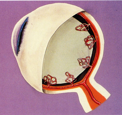
Blood sugar-related changes cause
new abnormal retinal blood vessels to grow.
Proliferative Diabetic Retinopathy (PDR)
Proliferative diabetic retinopathy is the most severe vision-related complication of diabetes, often leading to blindness if left untreated. It develops in 10 to 15 percent of diabetics.
Proliferative diabetic retinopathy begins in the same manner as background retinopathy but is then marked by the growth ("proliferation") of new blood vessels ("neovascularization") on the surface of the retina or optic nerve head. To further explain, with proliferative retinopathy, the retina is unable to obtain valuable nutrients needed for the vision process. To compensate, new blood vessels grow. Unfortunately, however, these vessels are very fragile and may end up further damaging the eye by causing other problems that can lead to permanent loss of vision and even loss of the eye, including:
Vitreous Hemorrhage: With this, the fragile new vessels rupture and bleed into the vitreous (the clear, jelly-like fluid that fills the center of the eye). Symptoms to watch for include fairly sudden loss or blurring of vision and seeing numerous floating spots (resembling spider webs) or a dense veil or dark spots.
Retinal Detachment: Although vitreous bleeding usually clears with time, it can (especially with repeated hemorrhages) create scar tissue. This scar tissue may tighten and pull on the retina, causing it to detach from the back of the eye. Symptoms include sudden onset of floaters or a cobweb or curtain effect. Retinal detachment may result in severe vision loss or even blindness.
Rubeosis: Growth of abnormal new blood vessels on the iris (the colored part of the eye), rather than the retina, that can lead to the following problem.
Neovascular Glaucoma: Rubeosis vessels on the iris can block the natural flow of fluid out of the eye. This increases pressure in the eye, leading to a severe form of glaucoma ("neovascular") that can result in permanent vision loss.
Proliferative diabetic retinopathy can occur in adult-onset diabetics, but it is more likely to develop in juvenile-onset diabetics, affecting almost 60 percent of those patients with more than 30 years of insulin-dependence. Over a five-year period, 50 percent of patients with the condition will become legally blind if they do not have treatment. Thus, it is especially important to detect this more severe form of diabetic retinopathy early.


Risk Factors
All diabetics (Type I or II, insulin-dependent or not) are at risk for developing retinopathy. Diabetes also doubles the risk for developing cataracts and glaucoma. The following factors can affect the incidence and degree of diabetic eye disease:
-
Time—The incidence and severity of retinopathy increases with the number of years you have diabetes. Patients with diabetes for less than five years have a 15 percent incidence of retinopathy. This skyrockets to about 70 percent after 10 years and 90 percent after 20 years.
-
Poor blood sugar control
-
High blood pressure
-
High cholesterol levels
-
Being overweight
-
Smoking
Symptoms
Background or Non-Proliferative Diabetic Retinopathy (NPDR)
Typically, there is no pain or redness, and symptoms include blurred or distorted vision of varying extent. Only when deposits or fluid involve the macular area is decreased vision actually noted. Thus, some patients with significant background diabetic retinopathy may still have excellent vision if the macula is spared.
Proliferative Diabetic Retinopathy (PDR)
For most people with proliferative retinopathy, there are no early symptoms. There is no pain, blurriness or inflammation. Patients may have good (or near normal) vision until the onset of vitreous hemorrhage or another serious problem causes symptoms. Others may, however, notice a change in their central and/or color vision as a result of macular edema (the leaking of blood vessels onto the central retina, or macula). This can often be treated if detected early.
Because of the lack of and/or variation in symptoms, it is vital for ALL diabetics to schedule regular comprehensive eye exams.

Detection & Monitoring
Diabetic eye disease is detected through comprehensive eye examinations involving dilation of the pupil (enlarging with drops) to best see into the back of the eye. The following methods may be used to diagnose and monitor diabetic retinopathy.
Ophthalmoscopy: An instrument called an ophthalmoscope is used to check for early signs of retinopathy, such as microaneurysms (tiny blister-like outcroppings on retinal blood vessels that can bulge and leak), before noticeable vision loss occurs.
Ultrasonic Testing: In cases where bleeding obscures viewing of the retina, ultrasonic testing can be useful to ensure that bleeding and scar tissue has not caused a retinal detachment.
Optical Coherence Tomography (OCT): Eye Care Specialists is pleased to join such prestigious centers across the country as Johns Hopkins and Bascom Palmer Eye Institute in offering the latest advancement in diabetic retinopathy diagnosis and tracking—Optical Coherence Tomography (OCT). Similar to a CT scan, the OCT creates detailed computer printouts that provide unparalleled accuracy in visualizing and measuring the severity and extent of swelling (“macular edema”) present from diabetic retinopathy.
During the fast, painless OCT procedure, patients simply focus on a light while a safe, invisible laser scans the back of the eye to acquire an image in just seconds. This “optical ultrasound” of the anatomic layers of the retina and optic nerve enables us to detect and track not only signs of diabetic retinopathy, but also glaucoma, macular degeneration, and other sight-threatening diseases—often before any damage occurs. (This is especially important for diabetics, since they are at a much greater risk for developing glaucoma and other vision problems.) Follow-up OCT scans are then used to promptly detect and treat any abnormalities with the necessary medications, laser therapy or surgery—thus helping to prevent any future loss of vision.
In addition to undergoing examination and testing, it is important that you provide your doctor with an accurate medical history, including any vision symptoms or concerns, hereditary problems, and medication use.

When to Schedule Exams
Do you have diabetes? Was your last eye exam more than a year ago?
If you can answer “Yes” to those two questions, then it’s time to have your vision checked. It is vital to understand that significant retinopathy may be present before noticeable vision loss occurs (if the macula is spared). As such, early detection and treatment are crucial to reducing the risk of vision loss from diabetes. Therefore, all diabetics should:
-
Schedule yearly comprehensive dilated eye exams (more often, if recommended).
-
Call your eye care specialist immediately if you notice floating spots (like spider webs), a "veil" over your vision, or other changes (not associated with blood sugar fluctuations).
-
Schedule an eye exam if you become pregnant, since this can speed the progression of retinopathy.
And, remember, whether you are diabetic or not, after age 40, EVERYONE should have their eyes checked at least every two years for other conditions, like glaucoma, that may be causing permanent vision loss without you even noticing it.
(NOTE: Eye appointments are often covered by insurance, Medicare or Medicaid; and payment and financing options are also often available.)

Controlling Diabetic Retinopathy
Although diabetic retinopathy can NOT be prevented or cured, prompt diagnosis, diligent monitoring and timely treatment can control the sight-robbing effects of diabetes. The following steps are used to protect vision.
Medical & Lifestyle Strategies
Blood Sugar Control
Besides being important for your general health, good long-term control of diabetes through medication, diet and exercise may also greatly reduce your risk of vision loss. Studies have shown:
-
In diabetics with no eye disease, excellent blood sugar control reduced the risk of developing retinopathy by 76%.
-
In patients with mild retinopathy, excellent blood sugar control slowed its progression by 54% and reduced the development of severe diabetic retinopathy by 47%.
Healthy Lifestyle
Proper diet and exercise are crucial to anyone's good health, especially people with diabetes. Also, controlling blood pressure and not smoking definitely decrease the complications of diabetes, including retinopathy.
Regular Check-Ups
See your doctor, endocrinologist and eye care specialist as often as recommended. Surprisingly, a study found that one-third of people who know they have diabetes do not adhere to guidelines for yearly dilated eye exams. Why do you need your eyes checked, especially if your blood sugar has always been under control? Regular eye exams are important because diabetes is also a disease of the tiny blood vessels, especially the capillaries, and retinopathy may progress independent of blood sugar levels. Duration of the disease (number of years having diabetes) is really the most critical factor in determining the development and extent of retinopathy and whether you need treatment.

Injection Treatment
Our practice has been very pleased with the success of new anti-VEGF medications (Avastin, Eylea, Lucentis and Vabysmo) that can be painlessly injected directly into the eye to decrease both blood vessel leakage and abnormal new growth—thus staving off progression of diabetic eye disease. (Click on video)
Our patients have experienced excellent results with injections, including not only stabilization of vision, but in some cases, measurable improvement in sight. Studies in 2011 established the primary role of medication injection in the treatment of diabetic macular edema. Protocols exist for the use and frequency of injections (typically every 4-12 weeks). Medication injections may also be used in conjunction with laser treatment, as described next.
Laser Treatment
Although not all diabetics can have or need laser treatment, it has proven very effective in reducing severe vision loss, especially if begun early enough. Its main goal, however, is to stabilize retinopathy—not to restore lost vision. Treatment is performed in our office or outpatient surgery center using topical (drops) or local (injection) anesthesia.
During laser photocoagulation, a high-energy beam of light is focused onto specific areas of the retina. This light turns to heat and seals leaking blood vessels (for background or non-proliferative retinopathy) or helps prevent the growth of abnormal new vessels (for proliferative retinopathy).
Surgical Treatment
If laser photocoagulation is impossible or unsuccessful in preventing recurrent vitreous hemorrhage, scar formation or retinal detachment, an operation known as a vitrectomy may be performed. This is a major surgical procedure in which the bloody fluid and abnormal scar tissue in the vitreous cavity are removed and replaced with a clear, artificial solution. Although complications may occur, about two-thirds of patients have some vision improvement with this technique.
Deciding when or whether to use injection, laser or surgical treatment depends on careful evaluation of the type, location and severity of your retinopathy and your doctor’s opinion as to how well the condition may (or does) respond to treatment.

Tips to Reduce Vision Loss
-
If you do NOT have diabetes, have a blood sugar test every three years after age 45 to screen for the disease.
-
If you DO have diabetes, check blood sugar levels regularly and hemoglobin A1c levels at least every four months. Aim for less than 7.0.
-
Keep blood glucose levels close to normal through diet, medication and exercise. (Fluctuations in blood sugar levels can temporarily affect vision, making it more difficult to detect serious problems. Good control slows the onset and progression of retinopathy and lessens the need for laser surgery.)
-
Keep blood pressure under control.
-
Don't smoke.
-
Keep cholesterol levels low.
-
Schedule dilated eye exams once a year, or as often as your eye care specialist suggests. Advances in diagnosis and treatment are of no value if you don't utilize them.
Blindness due to diabetes places physical, emotional and financial tolls on patients, their loved ones and society. Although, at present, we cannot prevent or cure diabetic retinopathy, early detection, careful tracking, and timely treatment can often decrease the risk of severe visual loss. Those are steps worth taking to gain every opportunity to see life to the fullest—now AND in the future.

For more information or a comprehensive examination . . .
Since 1985, Eye Care Specialists has provided comprehensive medical, surgical and laser care for virtually every eye condition to more than 200,000 people. If you would like our free educational booklet on the eye concern reviewed above, please complete this form or call our Communications & Education Department at 414-321-7520 ext. 235. To schedule a comprehensive eye examination or a second opinion evaluation, call any of our three convenient Milwaukee-area locations directly.